
Gross Anatomy
The liver is the largest gland in the body, with a reddish brown colour. The liver is protected by the thoracic cage and the diaphragm, lying beneath ribs 7 to 11 on the right side.
It has a diaphragmatic surface which is formed by the anterior, superior and some of the posterior surface. This is separated by the anterior inferior border from the visceral surface which is the inferior and some of the posterior surface of the liver. The diaphragmatic surface of the liver is smooth and dome shaped, allowing it to fit into the concave shape of the diaphragm. However it is separated from the diaphragm by a space called the subphrenic recess.
The visceral surface of the liver is related to many organs. It contacts the right side of the stomach, the gastric and pyloric areas. It is also related to the beginning of the duodeum, the gallbladder, the right kidney and supradrenal gland, the right colic flexure and right transverse colon. 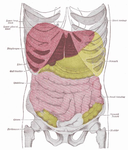
Figure: The Liver and its relations
Image courtesy of https://en.wikipedia.org/wiki/Image:Gray1224.png. This image is in the public domain and thus free of any copyright restrictions.
It is located below the diaphragm and separated into four lobes:
- Right
- Left
- Caudate
- Quadrate

The falciform ligament anteriorly separates the right and left lobe. The falciform ligament is a sheet of mesentery which suspends the liver from the diaphragm and the anterior abdominal wall, tethering it in place. The right lobe is further divided into the caudate and the quadrate lobe. Posteriorly the right and caudate lobes are separated by the deep groove formed by the inferior vena cava on the liver and boarded by the fissure which holds the ligamentum venosum.
Figure: The Anterior Liver
Image courtesy of https://upload.wikimedia.org/wikipedia/commons/b/ba/Gray1085.png. This image is in the public domain and thus free of any copyright restrictions.
It is also connected to the diaphragm and abdominal walls by the coronary, and the fibrous round ligament.
The round ligament is also called the ligamentum teres. It is a remainder of the umbilical vein. The umbilical vein carried nutrient rich, oxygenated blood from the placenta to the liver of the embryo.
The liver is completely covered by a layer of visceral peritoneum, except in the bare area. The bare area is formed by the coronary ligaments attachment to the diaphragm. The bare area is bounded by the two layers of the coronary ligament and the inferior vena cave. The coronary ligament edges are called the triangular ligaments. The porta hepatis and the bed of the gallbladder are also devoid of peritonium.
The caudate and the quadrate lobe are only present on the posterior of the liver. The caudate lobe is above the quadrate lobe and is next to the point of exit of the inferior vena cava from the liver. The quadrate lobe is below the caudate and sits next to the gall bladder. Between them is an opening called the porta hepatis.
Porta Hepatis
The Porta Hepatis is a transverse fissure on the visceral surface of the liver. It is where the hepatic portal vein and proper hepatic artery enter the liver, and where the bile ductules exit it. Collectively these 3 vessels are called the portal triad. Aswell as these vessels the porta hepatis allows passage of the hepatic nerve plexus, hepatic ducts and lymphatic vessels.
All the vessels travel in the lesser omentum which is a thin double layered peritoneum that extends from the liver to the stomach and the start of the duodenum.

Figure : Posterior Liver
Image courtesy of https://commons.wikimedia.org/wiki/Image:Gray1086-liver.PNG. This image is in the public domain and thus free of any copyright restrictions.
The gallbladder is situated in a depression on the posterior surface of the liver between the right and quadrate lobe. The gallbladder is a pear-shaped sac which stores bile synthesised by the liver.